AT A GLANCE
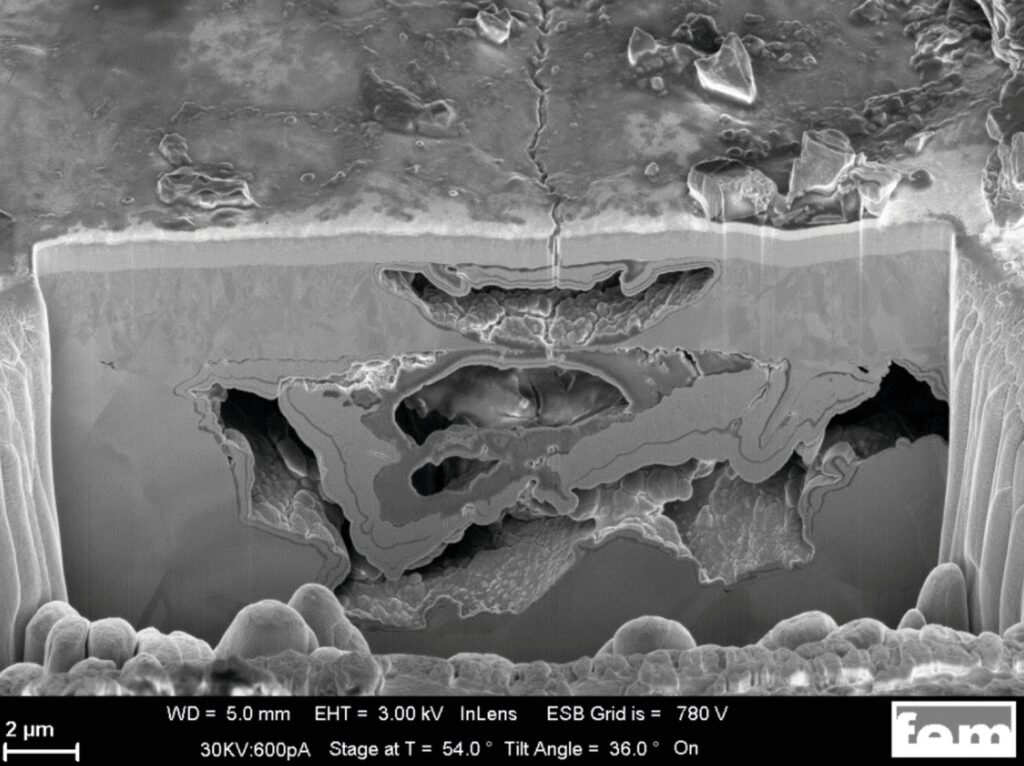
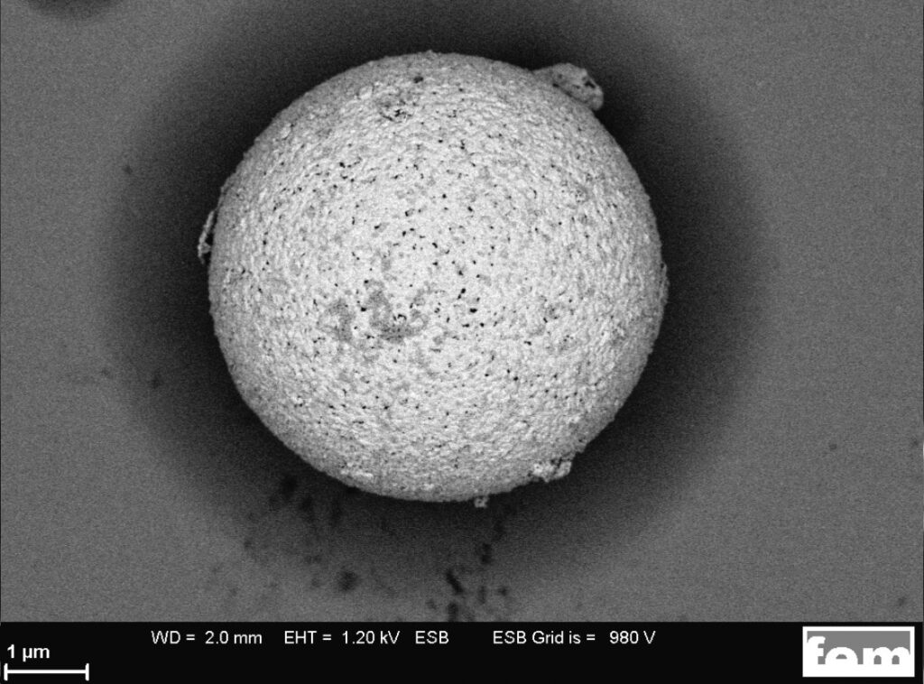
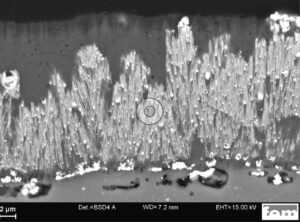
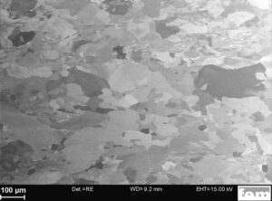
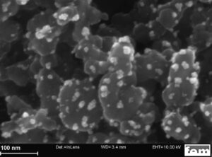
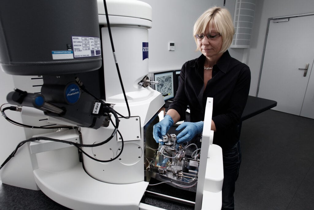
The scanning electron microscopes (SEM) equipped with a field emission cathode enable material analyses in the nanometre range. Images with the secondary electron (SE) detectors are topography or edge-emphasised, while images with the backscattered electron (RE) detector show the material or channeling contrast. Detectors for energy or wavelength dispersive X-ray spectrometry (EDX, WDX) are available for spatially resolved element analysis. The crystallographic properties can be analysed using electron backscatter diffraction (EBSD). EBSD provides information on phases, grain sizes and crystal orientations. The focussed ion beam (FIB) enables targeted material removal. With FIB, cross-sections in surfaces can be prepared with high spatial resolution and analysed in situ by electron microscopy. This method is therefore suitable for analysing layer systems, defects or corrosion, for example.
STRUCTURAL ANALYSES
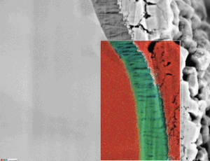
X-ray spectrometric element analysis